
i5dentalchennai@gmail.com
+91-9043765454
3D and 2D imaging services are important tools used in dentistry to help diagnose and plan treatments for various dental conditions. Here are some of the imaging services commonly used in dentistry:
Overall, 3D and 2D imaging services provide important diagnostic and planning tools for dentists to help improve patient outcomes and treatment results.
Cone beam computed tomography (CBCT) is a type of 3D imaging technology used in dentistry to provide detailed views of the teeth, jawbone, and other oral structures. CBCT uses a cone-shaped X-ray beam that rotates around the patient's head, capturing multiple images from different angles. These images are then reconstructed into a 3D image of the oral structures, allowing for a more detailed and accurate view compared to traditional 2D X-rays.
CBCT is commonly used in dentistry for implant planning, orthodontic treatment planning, and diagnosing jaw disorders. It can provide information about the quality and density of the jawbone, the position of the nerves and other vital structures, and the location of impacted teeth. CBCT is also useful in diagnosing and planning treatment for conditions such as TMJ disorders, sinus problems, and oral cancer.
While CBCT provides a more detailed view compared to traditional X-rays, it does expose patients to more radiation. However, the amount of radiation is still relatively low compared to medical CT scans, and the benefits of CBCT for diagnostic and treatment planning purposes often outweigh the risks. CBCT should only be used when necessary, and the amount of radiation exposure should always be minimized by using appropriate protocols and techniques.
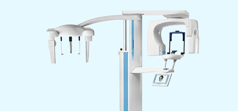
Digital X-rays are a type of 2D imaging technology that uses electronic sensors to capture and store images of the teeth and surrounding structures. Digital X-rays are a popular alternative to traditional film X-rays because they offer several advantages, including:
Digital X-rays are commonly used in dentistry for diagnosing dental conditions such as cavities, gum disease, and impacted teeth. They are also used for treatment planning, such as for orthodontic treatment or dental implants. Additionally, digital X-rays can be easily shared between dentists and specialists for consultation purposes.
Overall, digital X-rays provide several benefits over traditional film X-rays, including reduced radiation exposure, instant results, better image quality, and being more environmentally friendly.
Intraoral cameras are small cameras that are designed to capture images of the inside of the mouth. They are commonly used in dentistry as a diagnostic tool to help dentists identify and diagnose dental problems.
Intraoral cameras are typically handheld devices that are about the size of a dental mirror. They feature a small camera on the end that can be inserted into the mouth to capture images of the teeth, gums, and other oral structures. The images are then displayed on a computer screen or monitor, where the dentist can review them in real-time.
Intraoral cameras are commonly used in a variety of dental procedures, including routine check-ups, cleanings, and restorative procedures such as fillings and crowns. Overall, intraoral cameras are a valuable tool in modern dentistry, helping dentists provide more accurate diagnoses, improve patient education and communication, and increase efficiency.
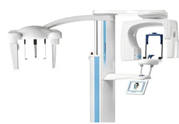
Panoramic X-rays, also known as panoramic radiographs, are a type of 2D dental imaging technology that captures a panoramic view of the entire mouth, including the teeth, jaws, and surrounding structures. Unlike traditional dental X-rays, which capture individual images of each tooth, a panoramic X-ray captures a single, wide-angle image that provides an overview of the entire oral cavity.
Panoramic X-rays are commonly used in a variety of dental procedures, including routine check-ups, orthodontic treatment planning, and implant placement. They are particularly useful for diagnosing complex dental problems and providing a comprehensive view of the entire oral cavity.
Overall, panoramic X-rays offer several benefits over traditional dental X-rays, including reduced radiation exposure, enhanced diagnostic capabilities, increased patient comfort, and time savings.
Digital models, also known as digital impressions, are a modern dental technology that allows dentists to create 3D digital images of a patient's teeth and gums. Digital models are created using a special scanner that captures detailed images of the teeth and gums, which are then used to create a virtual 3D model of the patient's mouth.
Digital models are commonly used in a variety of dental procedures, including orthodontic treatment planning, implant placement, and the creation of custom dental restorations such as crowns and bridges. They offer several benefits over traditional dental impressions, including increased accuracy, improved patient comfort, time savings, and enhanced treatment planning.
Lorem Ipsum is simply dummy text of the printing and typesetting industry. Lorem Ipsum has been the industry's standard dummy text ever since the 1500s


Lorem Ipsum has been the industry's standard dummy text ever since the 1500s, when an unknown printer took a galley of type and scrambled


Lorem Ipsum when an unknown printer took a galley of type and scrambled it to make a type specimen book. It has survived not only five centuries


Krishna Nagar, Pammal, Chennai
i5dentalchennai@gmail.com
+91-90437654540
© I5 Dental Implant Xperts. All Rights Reserved.
Designed & Developed By SBJ SERVICES